Thursday, November 11, 2010

The Future of Chiropractic?

I have decided to share my opinions with you tonight. After two wonderful speakers came into class today to discuss a complementary and alternative health department (that is administered by a Doctor of Chiropractic) in a local hospital, it led me to think about what the future of the chiropractic professional holds. There are several views and paradigms ranging from the strict upper cervical/neuro"philosophy" clinics (aptly named "Old School Docs" by the newer chiropractic generation) to those that I have previously mentioned who have integrated themselves into a hospital setting.

Patient care is number one. This is the general rule for every health care practitioner, whether it is a DPT (Doctor of Physical Therapy), Cardiothoracic Surgeon, or a homeopathic nurse, everyone unites under their concern for patient care. So explain to me, how or why wouldn't chiropractic want to jump on board? I cannot think of a single reason. Integrative medicine needs to become "good medicine" where the integrative approach is what is naturally and automatically implemented. There is a time and place for EVERY health care practitioner. I, as a soon-to-be Doctor of Chiropractic, will never claim to cure cancer, fix your cavities or even reverse your arthritis, I will, however, promise that I will serve you to the best of my abilities and give you and your needs the best clinical experience that I could possibly give, I refuse to let my ego get in the way of your care.

Chiropractic education has made dramatic leaps since the initial program implementations (Thanks Wilk! See previous posting about education hours, etc) and those who choose to embrace the general practitioner/primary care physician model are on the cutting edge of moving this profession to where it needs to be. At a recent graduation at my college, David Chapman-Smith (the secretary-general for the World Federation of Chiropractic) was quoted as saying the future of chiropractic lays within integration into sports rehabilitation and injury care, and I could not have said it better. Integration is key but not just in that one aspect. Sports injury care and on site evaluations will give chiropractic the leap forward that is needs. Luckily, I have been able to travel to a division 1 school to treat starting offensive linemen prior to games on several occasions but anyway...

These are what recent graduates are striving for every day: integrative practices, positively promoting chiropractic care, staying current on the neurological advances in why chiropractic works and what else we can do, staying current on research, and complete removal of the negative conceptions about chiropractic from skeptical minds.

Lack of compliance hinders progress. Ask questions! Be active in your health! Challenge your doctors, it will not only make them a better doctor but it will also make you more responsible for what's going on in your health!
Wednesday, November 10, 2010

Radiology #5 Synovial Chondromatosis

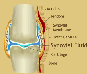
It has been quite awhile since I have updated my blog, so as I am sitting in clinic in between patient visits, I think I will take some time to educate the masses! This radiology blog will be about Synovial Chondromatosis also known as Synoviochondrometaplasia. Let's start out by explaining the joint capsule. Almost every joint in the body has a surrounding capsule. This capsule is lined with synovial tissue which secretes synovial fluid (lubrication for your joints, basically). Synovial chondrometaplasia is a condition in which there is proliferation of the synovial lining. The synovial lining proliferation can then calcify and break off, leaving a loose body (note: see arrows in the elbow radiograph) in the joint capsule.


There are two forms of synovial chondrometaplasia: primary and secondary. Primary synovial chondrometaplasia has no known onset or causation while the secondary form is usually due to increased joint stress or previous onset of osteoarthritis. Not only are there two forms, but there is a separate classification for how involved the joint capsule is, which correlates to the surgical treatment. Surgical treatment can range from a simple loose body removal or a complete synovectomy where the synovial lining is removed. Chiropractic management includes ultrasound and joint mobilization that can involve adjustments.

Sunday, July 4, 2010

Minimal Educational Requirements

Comparison of the Education of DCs and MDs
Both chiropractic and medical schools require certain course work for admission. These vary from school to school. Very few schools of either type require a bachelor's degree, although some specify that they prefer the applicant have such a degree.
Chiropractic colleges do not require the MCAT. Some medical schools do. Contrary to common belief, some medical schools (including high profile institutions) require the bare minimum of undergraduate requirements.
We took the admission requirements for medical schools from the publication titled: Medical School Admission Requirements, 1997-1998: United States and Canada, 47th edition (published by The Association of American Medical Colleges). Admission requirements for accredited chiropractic schools are dictated by the Council on Chiropractic Colleges (the agency appointed by the U.S. Dept. of Education to accredit chiropractic colleges).
The Parker College study reported that on average, chiropractic college involves 372 more classroom hours than medical school. Chiropractic students also have more hours of training in anatomy, physiology, diagnosis, and orthopedics (the musculoskeletal system).
It should be apparent from looking at the data below that in general, the chiropractic student has a more extensive classroom education and practical training in these areas, particularly in diagnosis, than the medical student.
Requirements for Admission to
Chiropractic and Medical Schools
College Courses
Parker Chiropractic College Harvard Medical School Stanford University
Biological Science (with lab) 1 year 1 year 1 year
General or Inorganic Chemistry 1 year 1 year 1 year
Organic Chemistry (with lab) 1 year 1 year 1 year
Physics (with lab) 1 year 1 year 1 year
English or Communicative Skills 1 year
Psychology 1/2 year
Humanities or Social Sciences 22.5 quarter hours
Electives 6-to-18 quarter hours.
Degree Requirements
These basic educational requirements for graduates of both chiropractic and medical schools show that although each has its own specialties, the hours of classroom instruction are about the same. (The class hours for basic science comparisons were compiled and averaged following a review of curricula of 18 chiropractic colleges and 22 medical schools.)
Minimum Required Hours
Chiropractic College Medical School
456 Anatomy/Embryology. 215
243 Physiology 174
296 Pathology 507
161 Chemistry/Biochemistry 100
145 Microbiology 145
408 Diagnosis 113
149 Neurology 171
56 Psychology/Psychiatry 323
66 Obstetrics & Gynecology 284
271 X-ray 13
168 Orthopedics 2
2,419 Total Hours for Degree 2,047

Friday, July 2, 2010

Radiology #4 Osteoarthritis

As I'm stuck in town for the holiday three day weekend so I can study for my upcoming diagnostic imaging III midterm, I decided to post about a topic that I'm going to be tested over. Let's first start off talking about arthritis. Everyone has seen the tylenol commercials for "joint pain" with the geriatric patients walking around holding their backs, necks, hand, etc anything to make the public sympathetic to a condition that they are already forming. Arthritis is defined as the inflammation of one or more joints which results in pain, stiffness and limited movement. There are over 100 different types of arthritis but today we will be focusing on osteoarthritis (OA) or as it's also known as degenerative joint disease. OA is the most common joint disorder, and it's not surprising because most injury sites, such as the fractures previously blogged about, will eventually turn into OA because they weren't properly rehabbed, patients didn't comply with doctor's orders, or simply from overuse. OA occurs when the cartilage lining the joints wears down, causing more stress on the bones in the joint. It's radiographically found as joint space narrowing with an increase in the cortical margins of the bones also known as subchondral sclerosis. Note the difference between the normal (top) knee radiograph and the one demonstrating OA (bottom). Medical treatment includes pain relievers and physical therapy. Chiropractic treatment includes adjusting to restore motion into the joint that has lost it's motion, physical therapy to rehab the surrounding musculature and ligaments and nutritional counseling, where applicable.

Thursday, July 1, 2010

Case #1 Inversion Ankle Sprain Injury

In an attempt to become more of an educator and less of an informative know-it-all, let me first start off this blog by talking about motion. Every joint in the human body has some sort of motion, whether it's pretending you can fly with your arms or tucking them under your pillow when you sleep. Many health care practitioners, including chiropractors, will, when a joint is injured or as a daily objective test will put a joint through it's entire range of motion. When the patient does this by themselves, it is known as active ranges of motion or AROM. Another test for ranges of motion include the patient relaxing the joint and letting the practitioner take the joint through it's motion, this is called passive ranges of motion or PROM (or as I tell my patients, I'm going to take you to your prom now!). If pain is provoked during PROM, this indicates ligamentous injury. (Ligaments attach bone to bone). The final range of motion testing is called resisted ranges of motion or RROM, where the practitioner will resist the patient as they actively put the joint in question through it's range of motion. If pain is provoked on RROM, the pain is due to muscular injury, as you are utilizing your muscles to resist the motion.

Onto the more interesting item at hand: Did you know that over 25,000 inversion ankle sprains are reported every day? Let me first define inversion ankle sprain. An inversion ankle sprain is when someone rolls their ankle in such a way that the bottom of their foot is facing the opposite foot or inward, compared to an eversion ankle sprain where the bottom of the foot is facing outward, away from the other foot. I currently have a young adult, male patient who came in with an extremely bruised and inflammed (swollen) left ankle. Mechanism of injury: he was playing lacrosse when he rolled his ankle via inversion and while rolling his ankle, he was pushed forward. After doing a thorough ankle regional examination, his PROM provoked pain which added a mark in my intial thought column of ligamentous injury (don't give me too much credit, it's how most inversion sprain injuries come out), orthopedic testing confirmed as well. If you will notice in the picture to the left of a normal ankle, there are three ligaments on the outside or lateral side of the ankle that attach the fibula (one of the lower leg long bones) to the ankle. The most common ligament to be injured is the anterior talofibular ligament (ATFL) which attaches the fibula to the talus, a tarsal bone that helps compose the ankle and ankle joint, as you can see there is a posterior one as well. Anterior and posterior refer to their location on the body, anterior basically means in front and posterior means in back. The second most common ligament to injure is the calcaneofibular ligament (CFL), which attaches the fibula to another tarsal bone in the foot, the calcaneus. The calcaneus is also the tarsal bone that makes up the heel of your foot. When inversion occurs in the ankle, the ligaments stretch out, this is clinically called a sprain. After taking my patient to the diagnostic ultrasound suite, my diagnoses had been confirmed. He had a grade 2 ATFL sprain and a grade 1 CFL sprain. Treatment for sprains include resting, icing, elevating, and compressing (RICE) the injuried area, usually via an ace bandage. We have been doing some cold laser therapy to the area which helps increase blood flow to the area which promotes a faster healing time, I'll post separately later about cold laser and it's clinical applications and research. We will eventually move to a more rehabilitative model to get his ankle back in shape, but as he has done this before, he will need to wear a brace or have his ankle taped every time he decides to participate in any sporting activity. Once ligaments are stretched out, it can take years (if ever) for them to return to normal size and shape.

Wednesday, June 30, 2010

Radiology #3 Radial & Ulnar Bone Fractures


Why hello! After a lovely lecture today about radial and ulnar bone (the two bones that make up your forearm) fractures in my diagnostic imaging class, and viewing an elbow series today in radiology conference, I've decided to post about four of the different fractures that occur and their mechanism of injury. The top radiograph is normal anatomy of the wrist, so that you may understand or more readily observe the fracture. The thicker of the two bones is radius, and the thinner of the two is the ulna, notice on the lateral projection (the xray on the right, the two bones are super imposed over each other, or line up). The first type of fracture is called a Colles' fracture. A Colles' fracture is a fracture of the distal (more towards the fingers) radius where the fracture fragment is displaced posteriorly. Colles' fractures are also classified as osteoporotic fractures and are usually the first place where a fracture appears in post-menopausal women. This type of fracture usually occurs when someone has fallen on (an) out stretched hand (FOOSH). The second type of fracture is a Smith's fracture aka reverse Colles' fracture where the fracture fragment is displaced anteriorly (palm side of the hand). This type occurs when someone doesn't have time to extend their wrist when falling, usually occurs in elderly patients whose mental capacity is fading. The third type of fracture is called a Galeazzi fracture. It's a fracture of the distal radius that is accompanied by an ulnar dislocation, this also often occurs from a FOOSH type injury, where the fracture radius is shortened. Notice that the "fracture fragment" remains with the carpal bones and the hand. Often radiologists will use the thumb as an indicator as to which direction the fracture will displace. The last type of fracture I'm going to discuss tonight is a Monteggia fracture, it's almost the opposite of a Galeazzi fracture. A monteggia fracture is fracture of the proximal ulna (towards the elbow) with a dislocation of the radius. The more serious of the last two types of fractures that I've listed are the dislocations because soft tissue healing is a longer, less developed process than bone healing. Some of the fractures, depending on the severity are surgical cases where screws, plates, bars, etc will be inserted and then casted. Can you say bad day?

Tuesday, June 29, 2010

Radiology #2 Stress Fractures

After a very overdue spin class, I decided to talk about stress fractures today, in the hopes that I do not acquire one. Stress fractures are caused by unusual or repetitive stress. Unusual stress may be someone deciding they are going to start training for a marathon, having never been a runner before in their life. Repetitive stress may be in the long term runner who overuses or pushes too hard in a short period of time. A radiograph is often negative for stress fractures within the first 4 weeks of injury but after those 4 weeks, callous formation and periosteal reactions are apparent. Let me define: callous meaning hard & thickened cortical bone (the thick, bright white lines outlining each bone), and periosteal meaning the membrane the covers the outside of the cortical bone. Case: 47 year old female who is a regular walker has been experiencing foot pain for several weeks now, the initial radiograph is negative for fracture.... as there are no breaks in any of the cortical lines. Four weeks later, the aforementioned patient has a follow up radiograph taken, where you notice the callous formation and periosteal reaction. Now it is important to take note that metastasis and infection can also cause periosteal reactions so patient history taking is VERY important in ruling in/out the more serious diagnoses. The second case is of a 37 year old female that complains of foot pain after a biking trip. The radiograph to the left is the initial, the middle is after 1 month, and the far right radiograph is a 3 month follow up. Treatment of stress fractures include: resting the injury site, usually a break from activities for 3+ weeks, icing and proper footwear evaluation.

Sources:

Berger, et al. Stress Fractures. Radiographical Department from the Academical Medical Centre, Amsterdam. http://www.radiologyassistant.nl/en/4615feaee7e0a 23 May 2007

Fractures without Significant Trauma.
Department of Radiology, University of Washington. 2007

Monday, June 28, 2010

Radiology #1 Scaphoid Bone Fracture

I have recently found a complete love for radiology! My college is lucky enough to have a brand new digital radiograph system, complete with a conference room where every day, almost all day long, the radiology interns and DACBRs (Doctors of Chiropractic who have a Diplomat from the American Chiropractic Board of Radiologists) read films, present case studies, etc. So from my love for that, not only will my radiology posts educate you, my reader, but will also affirm anything that I have learned, am learning or will learn. Since my cousin has a recent scaphoid bone fracture, I figured it would be the subject of my first radiographic post. The top radiograph (xray) shows normal anatomy of the wrist, and since the scaphoid is a carpal bone (one of the 8 small bones that makes up the wrist) a normal wrist radiograph is applicable. The pink dot is located adjacent to the scaphoid bone. You can clearly see the fracture in the next radiograph (note that these radiographs were taken on two different patients). Scaphoid fractures account for almost 71% of carpal fractures and are extremely unique due to it's vascularization. Usually a fracture of the scaphoid fracture will result in two pieces, like the one in this instance. The bone healing process is mainly dependent upon the blood supply (vascularity) because that's how nutrients arrive to the fracture site, hence healing. In the scaphoid, the blood supply is more proximal (closer to the wrist, farther from the fingers), so depending on where the fracture site is, the distal portion (the fracture piece closer to the fingers) may or may not get the nutrients that are vital to healing. Normal treatments include: casts over the wrist and may or may not include the thumb, surgical implementation of screws and bone grafts to adhere the fractured pieces together.

Sources:

Skeletal Trauma Radiology, University of Virginia. http://www.med-ed.virginia.edu/courses/rad/ext/

Boles, Carol. Wrist, Scaphoid Fractures and Complications. 22 March 2010; http://emedicine.medscape.com/article/397230-overview

Your Orthopedic Connection: Scaphoid Fractures of the Wrist May 2010. American Academy of Orthopedic Surgeons.

Hello Blogspot!


Chiropractic... what is chiropractic? As a politically active chiropractic student even I can say that this is controversial even withIN the profession. My definition of chiropractic is something that I have yet to completely compile, as it is constantly evolving. Chiropractors are the most qualified health care practitioners to take care of neuromusculoskeletal conditions, so yes this would be in my definition but chiropractic care is also applicable to ear infections, gastrointestinal problems, menstruation, etc the list goes on an on. I think everyone in the profession would agree that more research is needed but this thought also goes out to the pharmaceutical research. I would rather have quality research done at actual chiropractic colleges and from highly accredited medical colleges (example: Perceived benefit of Complementary and Alternative Medicine (CAM) for back pain: a national survey. Eisenberg, et al May 2010 JABFM) than studies that are FDA approved only because of the amount of money passing under the table. Chiropractic also falls under a wellness model of health care, and is actually the first profession (other than nutritionists) to fully integrate this into our treatment plans, philosophy and paradigms. If you think that you are 100% healthy because you have zero symptoms (whether it's pain, fever, headache, etc) then you are greatly mistaken.... but this is an entirely different topic altogether. So perhaps after this short discussion my definition of chiropractic thus far is that Chiropractic is a manual treatment therapy associated with treatment of the neuromusculoskeletal conditions and their somatovisceral correlations in the treatment of acute, chronic and wellness patients.